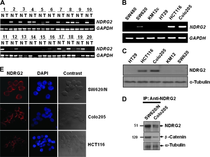
Fig. 1.
Differential expression of NDRG2 in colon tumor tissues and colon cancer cell lines. (A) NDRG2 mRNA levels in colon tumor tissues were evaluated by RT–PCR. T indicates colon tumor tissues and N represents normal mucosa tissues adjacent to tumor. Quality of total RNA used for RT was evaluated by gel electrophoresis on 1% denaturing agarose gel and the amount of RT products was normalized to glyceraldehyde 3-phosphate dehydrogenase (GAPDH). (B) Endogenous expression of NDRG2 in colon cancer cell lines was also examined by RT–PCR. (C) Expression level of NDRG2 protein in colon cancer cell lines was analyzed by western blot analysis. α-Tubulin was used as a loading control. (D) NDRG2 was immunoprecipitated with an anti-NDRG2 antibody, and the precipitant was analyzed by sodium dodecyl sulfate–polyacrylamide gel electrophoresis and western Blot analysis using anti-β-catenin antibody. (E) Intracellular localization of NDRG2 was determined using a confocal microscope. For nuclear staining, 4′,6-diamidino-2-phenylindole (DAPI) was used. NDGR2 introduced SW620 (SW620/N), HCT116 and Colo205 showed high expression levels of the protein, which localized mainly in the plasma membrane and cytosol.

