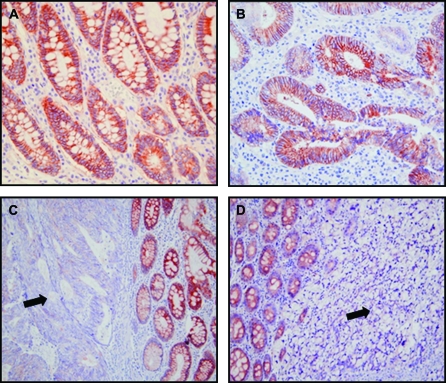
Fig. 2.
NDRG2 expression in patients with colon carcinoma. NDRG2 was highly expressed in the normal colonic mucosa, but its expression was decreased in colon cancer tissues as shown by immunohistochemical staining with anti-NDRG2 antibody. (A) Mucosal epithelial cells exhibited strong immunoreaction with a progressive increase along the crypt toward the upper part from the deeper part (×40 magnification). (B) In well-differentiated adenocarcinoma, NDRG2 was strongly expressed primarily on the cytosolic membrane of tumor cells (×40 magnification). The variations of staining in neoplastic cells correlated with the differentiation state of the tumor. (C) The staining signals in moderately differentiated adenocarcinomatous tissue (arrowhead) were very weak or null (×20 original magnification). (D) In poorly differentiated adenocarcinoma (arrowhead), tumor cells did not show any staining, whereas the adjacent normal epithelium showed strong signals (×20 original magnification).

