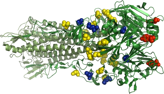
Figure 10. Locations of the predicted and confirmed stabilizing mutations to H1 hemagglutinin.
The full hemagglutinin trimer is shown in green, with the HA1 chains in dark green and the HA2 chains in light green. The temperature-sensitive mutation (ts-134 [104]–[106]) is shown with red spheres. The yellow spheres show the mutations that were predicted to be stabilizing by the PIPS program. The blue spheres show the four predicted mutations that were experimentally confirmed to actually increase the temperature stability. The structure is PDB code 1RVZ [107].

