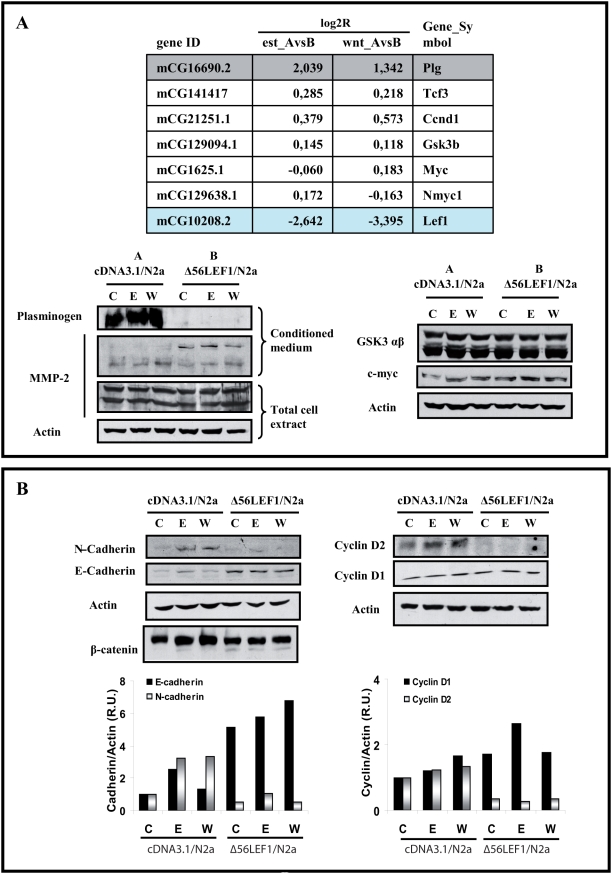
Figure 9. Gene expression of cDNA/N2a-m and Δ56LEF-1/N2a-m cells after exposure to estradiol.
(A)- Gene expression profile in cDNA/N2a-m and Δ56LEF-1/N2a-m stables cell lines. The upper panel reflects the gene induction of some selected genes, in microarray analysis of RNA collected after a 45 min exposure to estradiol or Wnt3a to detect the early response. Data is expressed as log2R from cDNA/N2a-m cells, that we denoted as group A, and Δ56LEF-1/N2a-m cells, that we denoted as group B. The effect of the treatment was compared between the two stable cell lines (A vs B) (see “Table 1” for a more complete list of the annotated genes). As seen, in the panel we selected some “putative Wnt-regulated genes”, such as Tcf3, Ccnd1 (cyclin D1), GSK3b, Myc and LEF-1, to give some examples of the results in our arrays. We detected changes at the protein level only in Plg, although there were several genes whose expression varied. For example, the levels of plasminogen RNA were much higher in group B than group A (ratio AvsB≥1), and the expression of LEF-1 was higher in Δ56LEF-1 due to the mutant expression (ratio AvsB≤−1). The western blots below are verifications of these differences at the protein level. Among other proteins that did not change between the groups of cells were GSK3 β or myc (see western blots on the right). MMP-2 was tested although it did not display a change in its RNA levels and there was no difference in the total cell extracts. Interestingly, when conditioned medium was prepared, more pro-active MMP-2 (and less active protein) was seen in Δ56LEF-1 cells. [Gene_Symbol: Plg (plasminogen), Tcf3, Ccnd1 (cyclin D1), GSK3b, Myc and LEF1]. (B)- Estradiol induction of N-cadherin and cyclin D2 may be affected by expression of the Δ56LEF-1 protein. Total cell extracts from cDNA/N2a-m cells (group A) and Δ56LEF-1/N2a-m cells (group B) were collected 24 h after estradiol or Wnt3a treatment to analyze several known Wnt or estrogen target genes. As seen in western blots, estradiol upregulated E-cadherin, N-cadherin and cyclin D2 expression in group A cells. N-cadherin and cyclin D2 were also upregulated by Wnt in group A cells. In contrast, E-cadherin expression was not Wnt responsive. The regulatory effects of estradiol on E-cadherin, N-cadherin and cyclin D2 expression were lost when Δ56LEF-1 is expressed, as seen in group B cells. In the case of E-cadherin, the loss of functional LEF-1, which acts as a known gene repressor, implies higher protein levels even without stimulation. In contrast, levels of actin or cyclin D1 remained unchanged.

