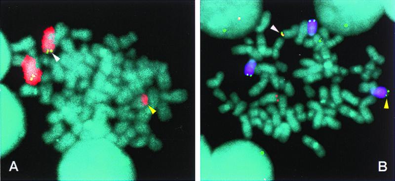
Figure 4.

FISH detection of a variant t(8;14) translocation in an MM tumor. Metaphase chromosomes from a primary MM tumor were hybridized with chromosome 8 painting (Texas red, red) and c-myc (FITC, green) probes (A) and chromosome 14 painting (Cy5, purple), CH (FITC, green), and c-myc (rhodamine, red) probes (B) to identify a variant t(8;14) translocation (diagram in Fig. 3A2). In both A and B, white arrows indicate the der(8) chromosome with c-myc and CH signals and yellow arrows indicate the der(14) chromosome that also has a CH signal. Because CH sequences are located on both der(8) and der(14), the chromosome 14 breakpoint appears to be located between the Eα1 and Eα2 3′ IgH enhancers, but could occur within the sequences detected by the CH probe or occur as a result of a duplication of CH sequences. In separate experiments not shown, a VH probe colocalized with the CH and c-myc probes on the der(8) chromosome (white arrows) but not with the CH signal on der(14).
