
Figure 2.
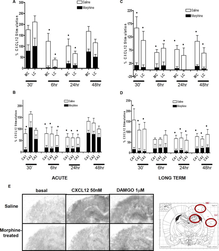
Morphine treatment reduces CXCL12-induced incorporation of [35S]GTPγS in the rat brain: pups were treated with saline or morphine acutely (ACUTE; single injection, 20 mg/kg, s.c.; A, B) or for an extended time period (LONG-TERM; once a day for 3 d, 10 mg/kg, s.c.; C, D) and killed after the indicated time. Brain slices were treated with CXCL12 (50 nm) and processed for GTPγS autoradiography. Representative autoradiograms of GTPγS incorporation after vehicle (left), CXCL12 (middle), and DAMGO (right) stimulation of either saline-treated or morphine-treated pups are reported below the graphs (E). Analysis was performed in different brain areas as indicated in the schematic diagram; data from medial and lateral cortex (MC and LC, respectively) are reported in the top graphs; the bottom graphs show data from the hippocampus (distinguished in field CA1, CA2, and CA3). Data are expressed as mean ± SEM of three animals per experimental group. (♦p < 0.01 saline treatment vs morphine treatment for each area reported in graph).