Fig. 1.
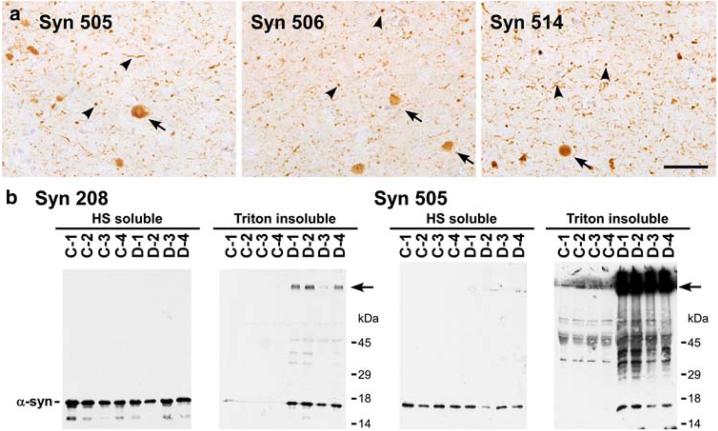
Immunohistochemistry and biochemical analysis of DLB brains with monoclonal antibodies that selectively detect pathological α-syn. a Immunostaining with Syn 505, Syn 506 and Syn 514 in the cingulate cortex of a DLB brain detecting LBs (arrows), but far greater neuritic inclusions and smaller “dot-like” aggregates (arrowheads) and relatively weak reactivity for the normal neuropil distribution of α-syn. (Bar scale = 50 μm). b Comparative immunoblotting analysis of biochemical brain samples using antibodies Syn 208 and Syn 505. The tissue from four controls (C1-4) and four DLB patients (D1-4) were fractionated biochemically as described in “Materials and methods.” HS-soluble and Triton-insoluble extracts were resolved on SDS-polyacrylamide gels and Western blots were probed with either Syn 208 or Syn 505. α-Syn was detected in the HS-fraction from all brains using either Syn 208 or Syn 505. Syn 208 detected monomeric α-syn predominantly in the Triton-insoluble extracts from DLB patients. Higher molecular mass α-syn aggregates, some that did not enter the resolving gel (arrow), were also weakly detected. Syn 505 also revealed Tritoninsoluble monomeric α-syn in diseased brains, but, in contrast, the major species detected were higher molecular mass α-syn aggregates that did not enter the resolving gel (arrow)

