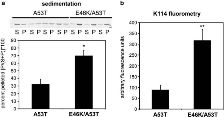
Fig. 4.
Analyses of the polymerization of A53T and E46K/A53T α-syn mutations. Recombinant A53T and E46K/A53T α-syn were incubated at 1 mg/ml for 72 h, as described in “Materials and methods.” a Representative Coomassie blue stained SDS-polyacrylamide showing the proteins in the soluble fractions (S) or in the pellets (P) following sedimentation analysis, and quantitative summary (* P = 0.003, n =7). b K114 amyloid fluorometry analyses (** P = 0.001, n = 7) demonstrating the increased propensity of E46K/A53T α-syn to polymerize compared to A53T α-syn. Data represent averages ± S.E.M

