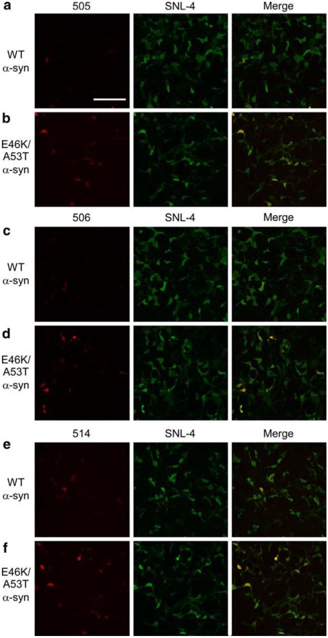
Fig. 5.
Double-immunofluorescence analysis of QBI293 cells transfected with WT or E46K/A53T α-syn. Representative fields of QBI293 cells transfected with WT α-syn (a, c, d) or E46K/A53T α-syn (b, d, f). Cells were immunostained with either Syn 505 (a, b), Syn 506 (c, d), or Syn 514 (e, f) (each in red, left) and with SNL-4 (green, center). Increased single-label immunofluorescence (left panels) and double-label immunofluorescence (merge, right panels) with Syn 505, Syn 506, and Syn 514 were noted in E46K/A53T α-syn over WT α-syn transfected cells. Representative images were chosen from a single experiment. (Bar scale = 200 μm)

