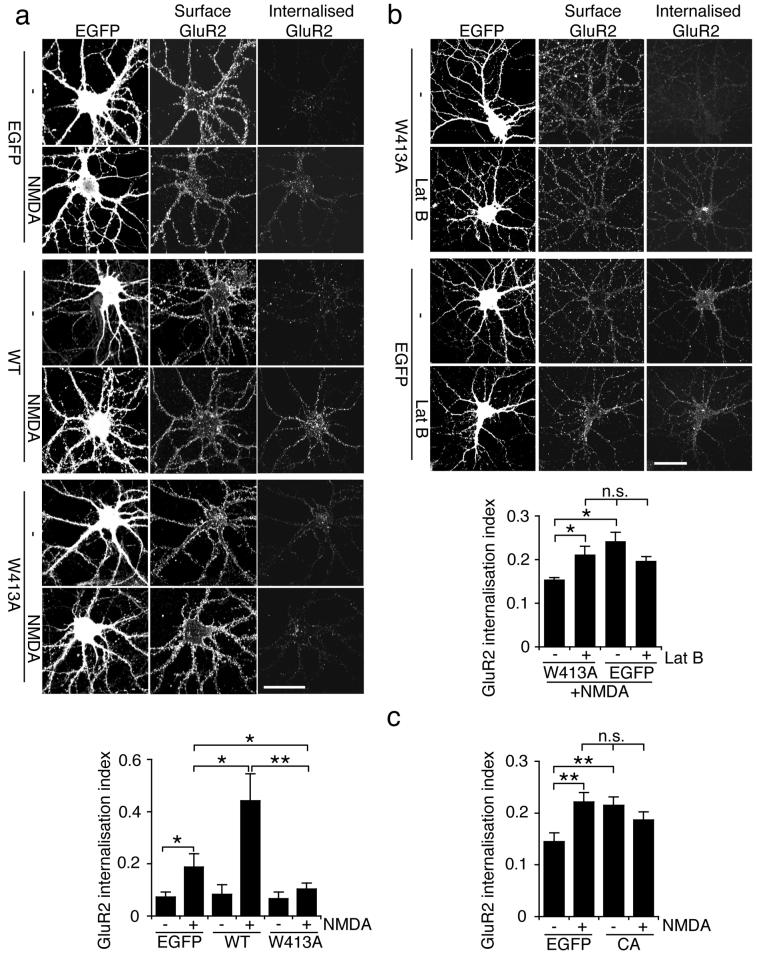
Figure 6.
Inhibition of Arp2/3-mediated actin polymerisation by PICK1 is required for NMDA-induced AMPAR endocytosis in neurons.
(a) W413A-PICK1 overexpression blocks NMDA-induced internalisation of GluR2-containing AMPARs. Dissociated hippocampal neurons were transduced with either WT-PICK1-IRES-EGFP, W413A-PICK1-IRES-EGFP, or empty-IRES-EGFP Sindbis virus. Internalised GluR2 in response to NMDA treatment was analysed by antibody-feeding using anti-GluR2. Cells were fixed and subsequently stained for surface and internalised GluR2 using different fluorescent secondary antibodies. Representative images are shown for all conditions, and graph shows GluR2 internalisation index (internalised/surface+internalised). n=12 cells, *p<0.05; **p<0.01. Data are means +/− SEM. Scale bar: 20 μm.
(b) Latrunculin B reverses the blockade of NMDA-induced AMPAR endocytosis by W413A-PICK1. Dissociated hippocampal neurons transduced with W413A-PICK1-IRES-EGFP or empty-IRES-EGFP Sindbis virus were exposed to 20 μM Lat B or vehicle for 1h, and treated with NMDA. Note all cells are NMDA-treated in this experiment. Representative images are shown for all conditions, and graph shows GluR2 internalisation index (internalised/surface+internalised). n=25 cells, *p<0.05, n.s. p>0.05. Data are means +/− SEM. Scale bar: 20 μm.
(c) Arp2/3 inhibition by N-WASP CA domain enhances AMPAR endocytosis. Dissociated hippocampal neurons were transfected with N-WASP CA-IRES-EGFP or empty-IRES-EGFP. Internalised GluR2 in response to NMDA treatment was analysed by antibody-feeding using anti-GluR2. Cells were fixed and subsequently stained for surface and internalised GluR2 using different fluorescent secondary antibodies. Graph shows GluR2 internalisation index (internalised/surface+internalised). n=20 cells, **p<0.01. Data are means +/− SEM.

