
Fig. 8.
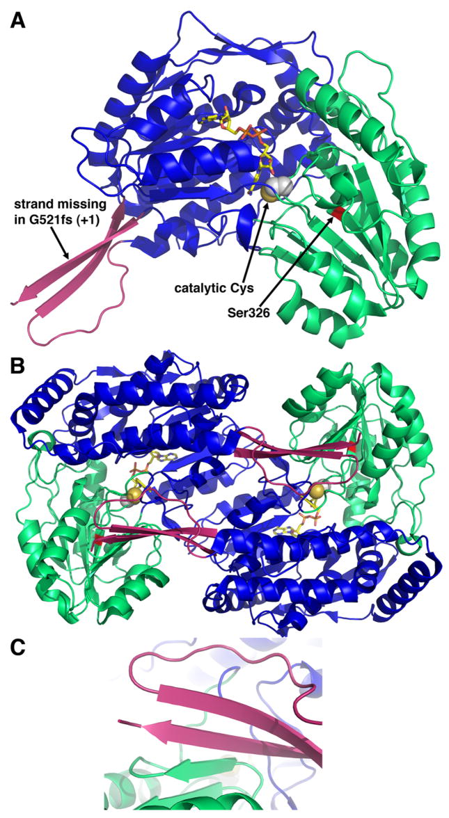
Structure P5CDH from T. thermophilus (Inagaki et al. 2006). A, ribbon drawing of a P5CDH subunit. The three domains are colored blue (NAD+-binding), green (catalytic) and pink (dimerization). The NAD+ cofactor is shown in yellow sticks. Catalytic Cys322 is represented in spheres. B, ribbon drawing of a P5CDH dimer. The two subunits of the dimer are colored as in panel A. C, close-up view of the intermolecular β-sheet in the dimer interface.