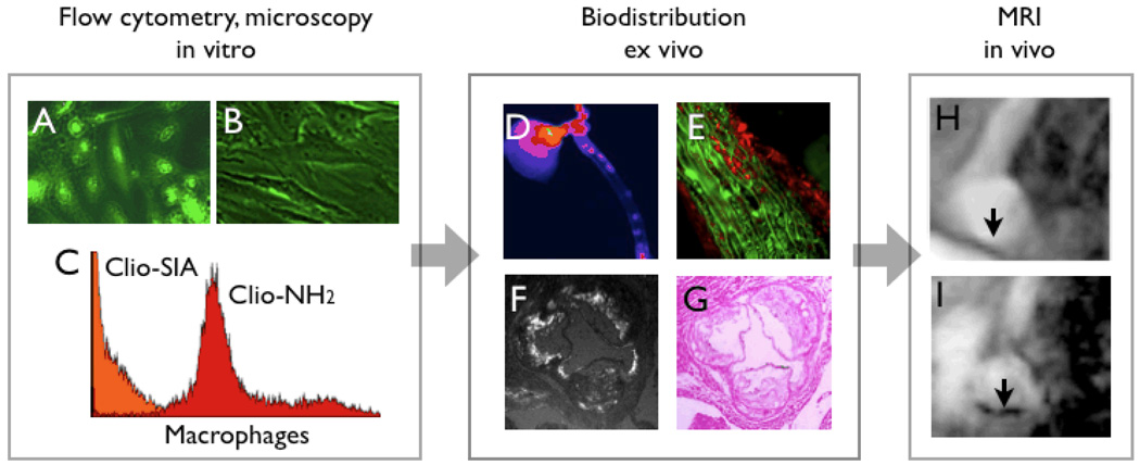
Figure 1. Role of NIR fluorochromes in agent development and validation.
1A & B: fluorescence microscopy of endothelial cells which have been incubated with a targeted magnetofluorescent nanoparticle (A). The uptake can be competetively inhibited (B), which demonstrates specificity of a candidate agent.
1C: Incubation of murine peritoneal macrophages with magnetofluorescent nanoparticles with different surface modifications. Through small molecule surface modifications, it is possible to completely re-direct MNP from macrophages (red) to endothelial cells (orange).
1D: Fluorescence reflectance imaging of an excised aorta, after injection of magnetofluorescent nanoparticles into a mouse model with atherosclerosis. This approach allows for rapid and cost-effective analysis of agent performance.
1E–F: Fluorescent properties of nanoparticles allow for microscopic analysis of uptake. In 1E, uptake of the nanoparticle (red channel in fluorescence microscopy) into smooth muscle cells (green, immunoreactive staining for alpha actin) of the media underneath a plaque is shown. F & G demonstrate uptake into plaques in the aortic root of an apoE−/− mouse.
1H&I: Finally, more time- and costintensive in vivo vascular imaging demonstrates uptake of nanoparticles in T2* weighted gradient echo imaging of the aortic arch (I) [15].

