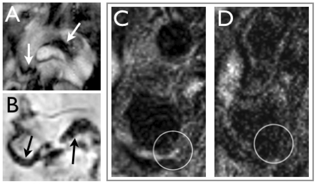
Figure 2. Macrophage imaging with magnetic nanoparticles.
2A: In vivo bright-blood aortic MR images of representative apoE−/− mice. Twenty-four hours following 15 mg Fe/kg of MNP injection, T2*-weighted gradient echo imaging was performed in a 9.4-T MR scanner.
Strong focal signal loss was observed in the aortic root and transverse aortic arch (arrows) [15].
2B: Ex vivo 14-T high-resolution aortic MRI of representative apoE−/− mouse that received MNP. Focal MRI signal loss was noted in the aortic root and transverse arch, similar to the in vivo images (arrows) [15].
2C&D: MRI of a carotid artery containing atherosclerotic plaques (circle) before (C) and after injection of MNP (D) in a patient [18].

