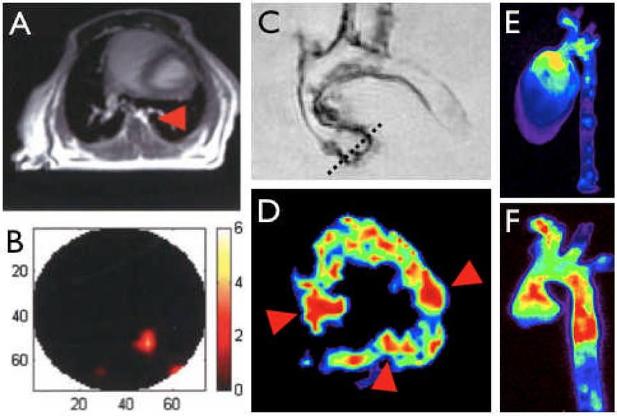
Figure 4. Hybrid imaging.
4A&B: Image co-registration using MRI and FMT combine the excellent anatomical detail in MR imaging (A, arowhead depicts descending thoracic aorta) with the sensitive detection of optical reporters in FMT [41].
4C–F: Combination of multiple molecular imaging agents in consecutive sessions or simultaneous imaging enable interrogation of complex biological systems. In C, an excised aorta from an apoE−/− mouse injected with a VCAM-1 targeted MNP is shown. The dotted line outlines slice positioning for color coded T2 weighted short axis MRI of the aortic root (D), which shows enhanced uptake in the valve commissures (arrowheads). E & F show fluorescence reflectance imaging after injection of a macrophage-avid nanoparticle and a protease activatable agent [1].

