
Figure 5.
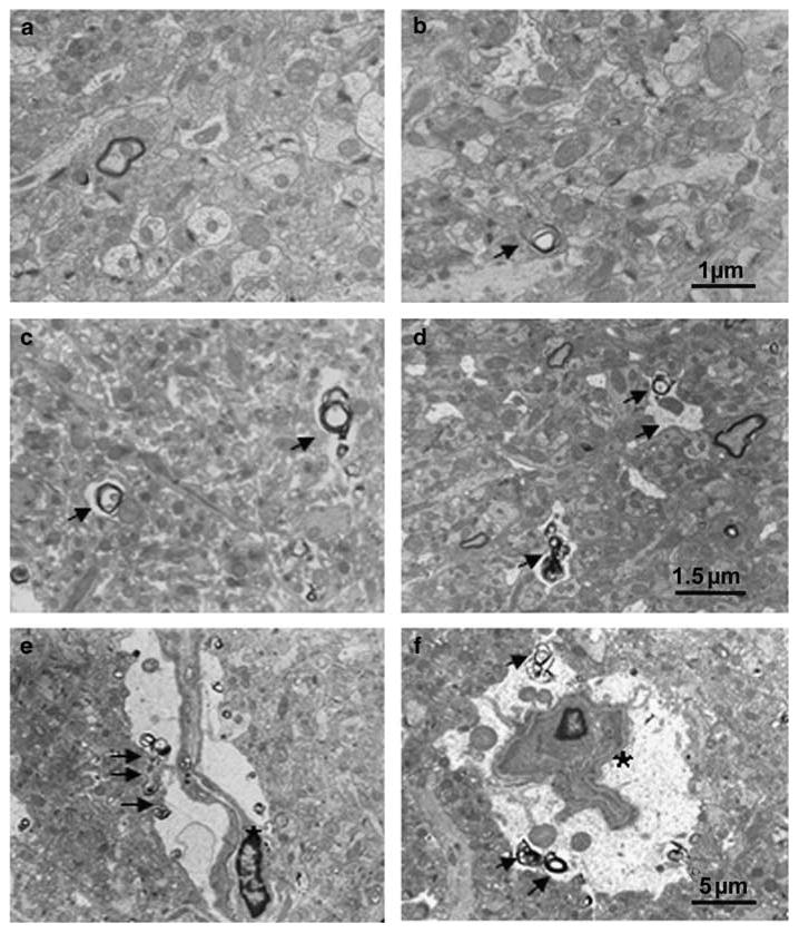
Electron microscope photographs taken in the molecular layer of the dentate gyrus following 6 months of memantine administration. (panel a) Tg + mice treated with vehicle showed decreased synapse density, but no degenerating axonal terminals. (panel b) Tg + mice with 5 mg/kg memantine demonstrated an increased synaptic density as compared to Tg + mice treated with vehicle (a), but no remarkable degenerating axons were observed. (Panel c and d) Tg + mice treated with 10 mg/kg (c) and 20 mg/kg (d) of memantine show increased degenerating axonal terminals (arrows indicated). Degenerating glial cells showing cell shrinkage with irregular space between perikarya and around neuropil (panel e and f, asterisks indicated). Bar in b applies to a; Bar in d applies to c; Bar in f applies to e.