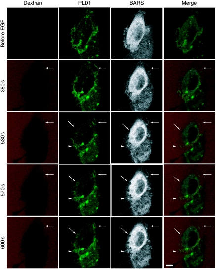
Figure 8.
Involvement of both PLD1 and CtBP1/BARS in EGF-induced macropinocytosis. COS7 cells were transiently co-transfected with expression vectors encoding GFP–PLD1 or CFP–CtBP1/BARS and cultured for 2 days. Cells were serum-starved for 1 h and stimulated with 100 ng/ml EGF in the presence of tetramethylrhodamine-labelled dextran and analysed for macropinocytosis by confocal microscopy. Representative frames of time-lapse images for GFP–PLD1 (green), CFP–CtBP1/BARS (grey) and tetramethylrhodamine-labelled dextran (red) and merged signals are shown. Arrows and arrowheads indicate newly forming macropinosomes. Bars, 10 μm.

