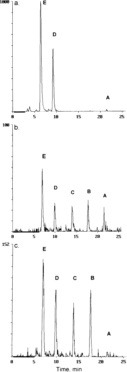
FIG. 3.
Representative HPLC radiochemical detector traces showing MXC and metabolites in (a) bile; (b) extract of liver; and (c) extract of intestinal mucosa. The chromatography was performed on a C18 column under conditions described in Table 2. Peaks are as follows: A, MXC; B, OH-MXC; C, HPTE; D, OH-MXC-glucuronide; E, HPTE-glucuronide. Similar chromatograms were obtained with other samples.

