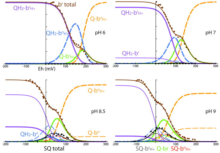
Figure 8.
Redox titrations at several different pHs for heme bH (brown) and SQi (black) in Rb. sphaeroides membranes. These fits do not assume that SQi is EPR silent when heme bH is oxidized. With three redox states of Qi and two redox states of heme bH, each of which can be protonated or not, there are in principle 12 possible species in this system; however, in practice several minor species can be ignored. Protonated states of heme bH are shown as thick lines, unprotonated as thin; reduced states of heme bH are shown as solid lines, oxidized states as dashed lines. Optical redox titrations of heme bH reported here are shown as brown dots, with the brown fit line showing the total b reduced. EPR redox titrations of semiquinone (Robertson et al., 1984) are shown as black dots, with the black fit line the total SQi.

