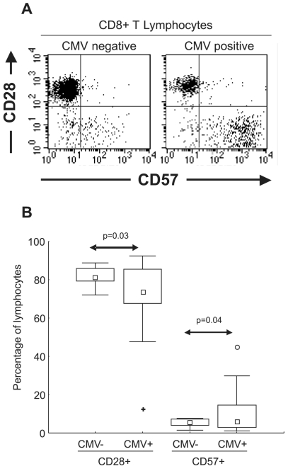
Figure 1.
(A) Graphic representation of CD8+ T lymphocytes (using CD3+CD8+ dual staining over a lymphocyte gate) from CMV seronegative and CMV seropositive healthy subjects. Expression of CD28 and CD57 were essentially mutually exclusive. (B) Distribution of CD28 and CD57 in 9 CMV seronegative (CMV−) and 24 CMV seropositive (CMV+) healthy subjects. Statistically significant differences were observed in the distribution of both subsets correlating with CMV serostatus (arrows) □ Median; □ 25%–75%; ⊤ Non-Outlier Min-Max; ○ Outliers; ✚ Extremes

