
Figure 1.
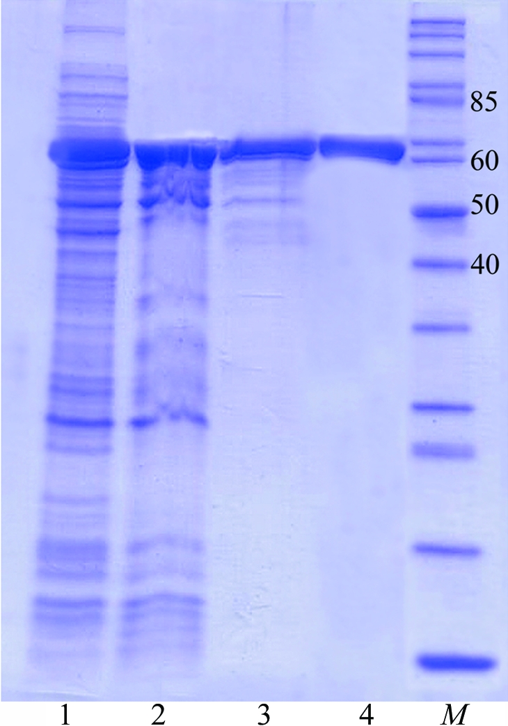
SDS–PAGE analysis of LigTh1519 during purification. Proteins were analysed on 12%(w/v) SDS–PAGE and were stained with Coomassie Blue. Lane 1, supernatant after removal of cell debris. Lane 2, supernatant after heat-treatment at 338 K. Lane 3, LigTh1519 after Ni-Sepharose FF column chromatography. Lane 4, purified LigTh1519 after gel filtration. Lane M, molecular-weight markers (kDa).