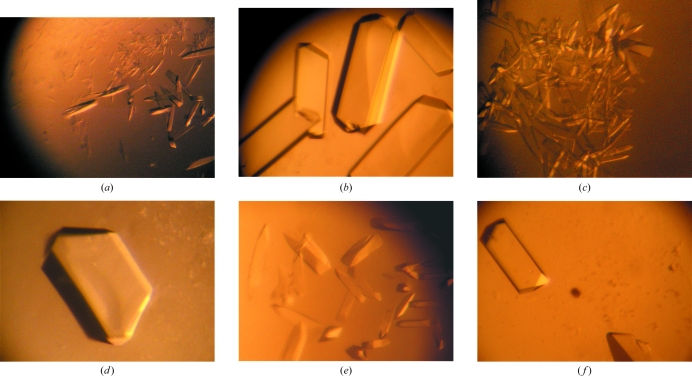
Figure 3.
Crystals of ShuA (a, c, e), ShuA-Pb (b), ShuA-Tb (d) and ShuA-Eu (f). All crystals were obtained using either 13–15% PEG 1K (a, b), 11–14% PEG 1.5K (c, d) or 8–12% PEG 3350 (e, f) with 0.1 M MES pH 6.5 and 0.1 M NaCl. The protein concentration was 10 mg ml−1 in 10 mM Tris–HCl pH 8.0 with 1.4% octyl-β-d-glucopyranoside. The heavy-atom concentration in the protein solution was 1.4 mM.

