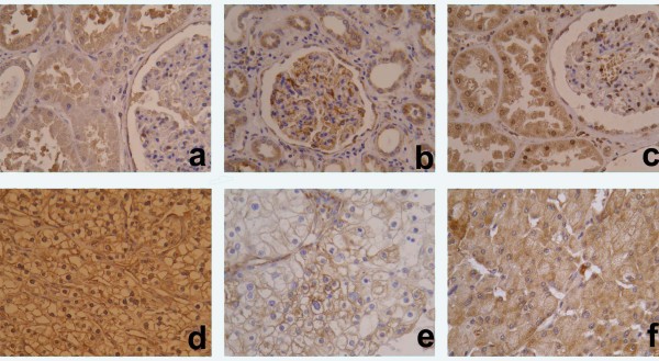
Figure 1.
Immunohistochemical staining of HIF-1α, VEGF-A and VEGF-C in normal renal tissue (A-C) and clear cell renal cell carcinoma (CCRCC) (D-F). A homogeneous cytoplasmic staining of tubular cells and weak staining in glomerules was observed with HIF-1α (A), while VEGF-A and VEGF-C were positive in tubular cells, glomerular mesangium and interstitial macrophages (B and C). In CCRCC, HIF-1α immmunoreactivity was nuclear and/or cytoplasmic (D), while it was perimembranous and/or diffuse cytoplasmic for VEGF-A and VEFG-C (E and F). (magnification ×200).

