Figure 4.

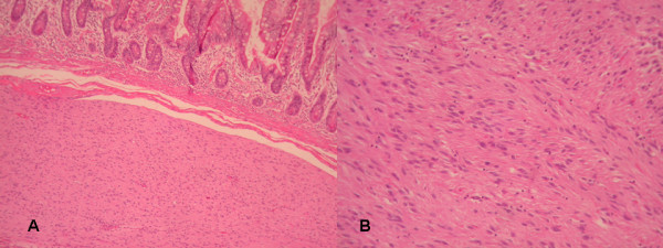
A) Invasion of submucosa of small intestine from GIST (HE×200). B) Histologically, the GIST was composed of sheets of spindle cell with moderate to slight interstitial collagen (HE×200).

A) Invasion of submucosa of small intestine from GIST (HE×200). B) Histologically, the GIST was composed of sheets of spindle cell with moderate to slight interstitial collagen (HE×200).