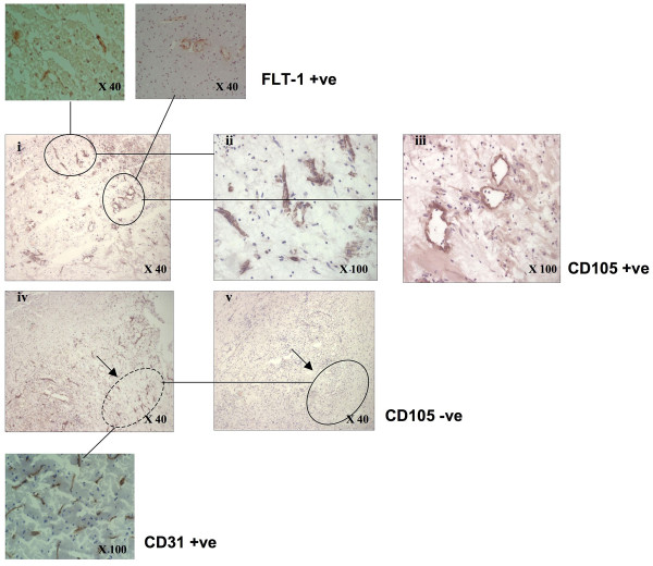
Figure 1.
Photomicrograph showing CD105-positive microvessels in histological areas chosen for laser-capture microvessels in peri-infarcted brain tissue (i-iii). CD105-positive clusters of blood vessels (inserts-top show the vessels were also Flt-1-positive. (iv) CD31-positive area (circled; insert) and (v) this area stained negative for CD105 (circle).

