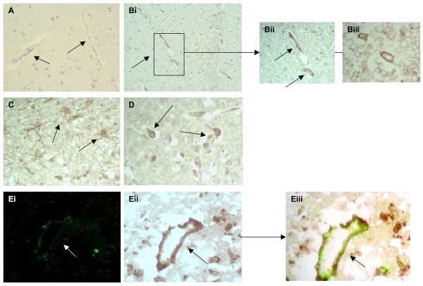
Figure 6.
MMP-2 was expressed strongly in stroked regions: A, tissue from the contralateral hemisphere showed no observable staining (negatively stained blood vessels are marked with arrows; × 40). B, peri-infarcted stroked brain tissue showing microvessels stained positive for MMP-2 (I; × 40; arrows) and (ii) at higher magnification (× 100) and (iii) in stroke/infarcted tissue (× 100; arrows). C, shows positive staining of cells with the morphological appearance of astrocytes/glia staining for MMP-2 in infarcted tissue (× 100; arrows) and D, cytoplasmic staining of neurones in the same region (× 100; arrows). Ei-iii, double immunoflourescence showing co-localization of MMP-2 (red) and CD105 (green) in peri-infarcted stroke tissue (× 100; arrow; sections from I0715P were used).

