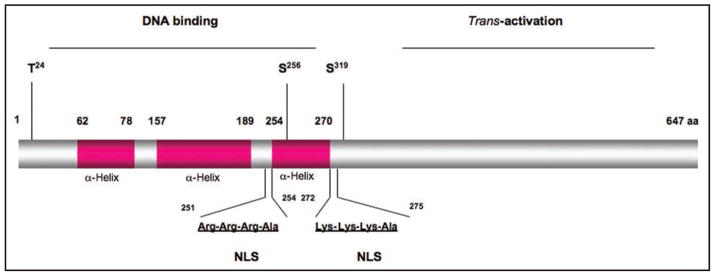
Figure 4.
Schematic depiction of FoxO1 protein. FoxO1 comprises the amino DNA binding domain and carboxyl trans-activation domain. The DNA binding domain is formed by three α-helix structural motifs. Within the DNA binding domain are two consensus nuclear localization signals (NLS) and three highly conserved phosphorylation sties (T24, S256 and S319). Phosphorylation of FoxO1 in response to insulin promotes FoxO1 translocation from the nucleus to cytoplasm. This effect results in inactivation of FoxO1 transcriptional activity and inhibition of target gene expression, as FoxO1 is removed from its active nuclear location.

