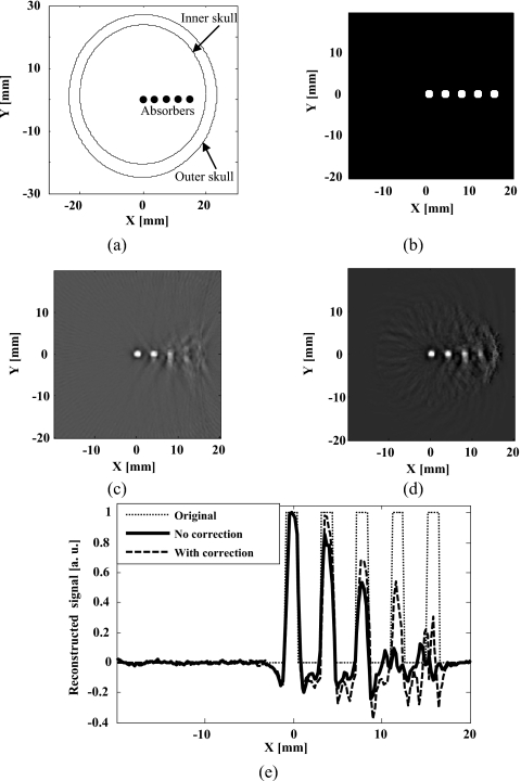
Figure 3.
Numerical simulation: (a) Schematic illustration of the phantom sample used in the simulation; (b) close-up view of the five absorbers, shown as white spots in the imaging area; (c) reconstructed TAT image without correction for skull effects; (d) reconstructed image after correction for skull effects; and (e) comparison of the reconstructed signals across the five absorbers.

