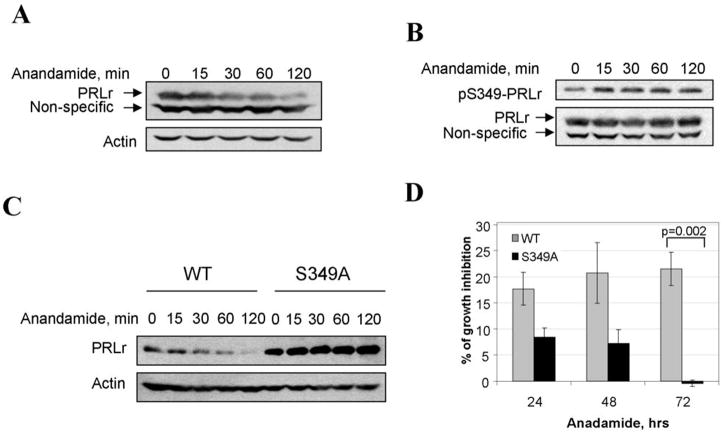
Figure 4. Anandamide promotes phosphorylation and downregulation of PRLr and inhibits growth of human breast cells.
A. Effect of anandamide on PRLr expression in T47D cells. T47D cells were treated with 10μM of anandamide and harvested at indicated times. The lysates (100μg of protein) were separated on SDS PAGE and analyzed by immunoblotting with antibodies against PRLr (H-300,) upper panels; and Actin (Affinity BioReagents) for loading control, bottom panels.
B. Effect of anandamide on S349-PRLr phosphorylation in T47D cells. T47D cells were pretreated with 20mM of methylamine for 2 hours, and then with 10μM of anandamide for indicated times. The lysates (100μg of protein) were separated on SDS PAGE and analyzed by immunoblotting with antibodies against pSer349-PRLr upper panel; and PRLr, bottom panel.
C. Effect of anandamide on PRLr expression in 53-MCF10A cells. 53-MCF10A stable cell lines expressing flag-tagged wild type PRLr (WT) or S349A mutant (S349A) were treated with 10μM of anandamide and harvested at indicated times. The lysates (100μg of protein) were separated on SDS PAGE and analyzed by immunoblotting with antibodies against Flag, upper panels; and Actin for loading control, bottom panels.
D. Effect of anandamide on 53-MCF10A cell growth. The graph represents the percent of the difference in the number of cells between ethanol and Anandamide treated groups ± SE at indicated times after the treatment calculated as described in the Materials and Methods. The differences between groups were statistical significant (p < 0.01) at 72h after the cell seeding.

