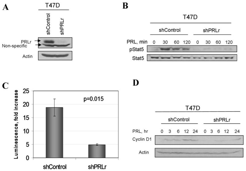
Figure 5. Knockdown of PRLr inhibits PRL signaling in T47D cells.
A. Expression of PRLr in T47D cells transduced with indicated shRNA constructs was analyzed by immunoblotting using indicated antibodies (upper panel). Analysis of β-actin levels was used as a loading control (lower panel).<br>B. Phosphorylation and levels of Stat5 proteins in T47D cell lines treated with PRL (100ng/ml) for indicated time were analyzed using indicated antibodies.
C. PRL-induced CISH promoter-driven luciferase activity in indicated T47D cell lines was performed as described in the Materials and Methods.
D. Levels of cyclin D1 in indicated T47D cell lines treated with PRL for indicated times were analyzed by immunoblotting. Same assay using anti-β-actin antibody is used as a loading control.

