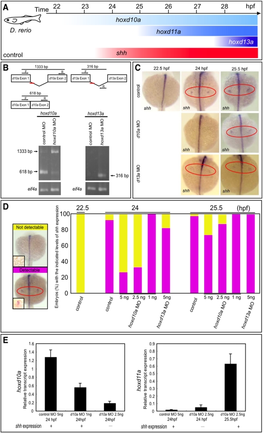
Figure 2. Timing of shh expression in zebrafish embryo fin primordia depends on hox transcript accumulation.
(A) Schematic representation of temporal hox and shh expression in the pectoral fin primordia of zebrafish embryos. shh was expressed at 24 hpf concomitantly with hoxd10a expression. (B) RT-PCR analysis to determine the efficiency of the hoxd10a or hoxd13a splice-blocking morpholino (MO). In the schematics, arrows represent forward (F) and reverse (R) primers, and the short red bars represent the hoxd10a MO and hoxd13a MO. Lower panel, analysis of RT-PCR products by agarose gel electrophoresis. Products of 618 bp and 1333 bp represent spliced and unspliced hoxd10a mRNA, respectively. The 316-bp RT-PCR product represents spliced hoxd13a mRNA. Amplification of eif4a cDNA was used as a control. (C) Whole-mount in situ hybridization to detect shh expression in the pectoral fin primordia of D. rerio embryos injected with 5 ng control MO (top panels), 5 ng hoxd10a MO (middle panels) or 5 ng hoxd13a MO (bottom panels) at the indicated hpf. Red ovals highlight the pectoral fin primordia. Note that shh expression was first observed at 24 hpf in the fin primordia of embryos injected with control (top) or hoxd13a MO (bottom), whereas shh transcripts became detectable at 25.5 hpf in the primordia of most embryos injected with hoxd10a MO (middle). (D) Percentages of embryos with detectable or undetectable levels of shh expression observed at 22.5, 24, and 25.5 hpf following injection of control MO, hoxd10a MO or hoxd13a MO (see also Figure S4). A representative image depicting the detectable or undetectable levels of shh expression in the pectoral fin primordia is shown at the left. Insets show high magnification views of pectoral fin primordia. (E) Semi-quantitative RT-PCR analysis to determine the expression levels of 5′ hoxd when shh is transcribed in pectoral fin buds. The relative levels of hoxd10a and hoxd11a transcripts in the lateral plate mesoderm of morphants were quantified. Relative expression was normalized against gapdh transcripts.

