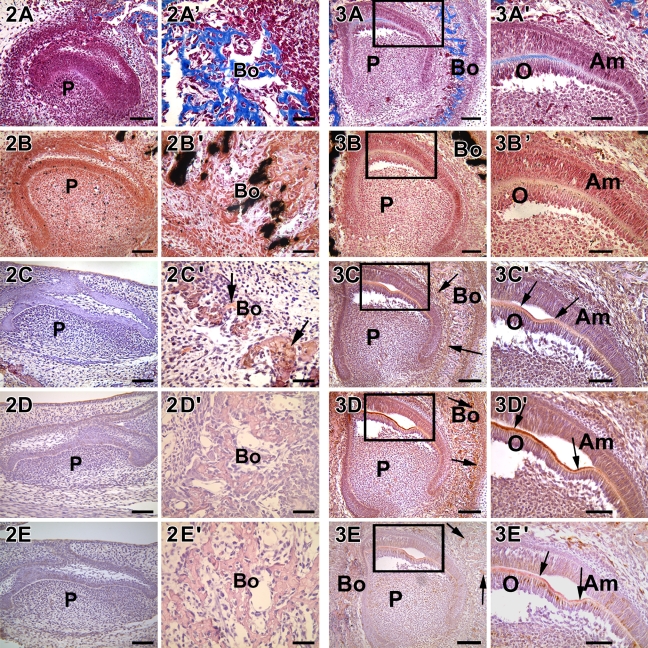
Figure 2.
Localization of collagen, mineral deposits, DMP1, DSP, and DPP in 18-day-old mouse embryos. Developing heads were collected from E18.5 mouse embryo, fixed, embedded, and cut into 5-μm-thick sections. DMP1 was expressed only in the osteoblasts of alveolar bone (C′, arrows). DPP and DSP were not expressed at this stage of development. Presence of collagenous matrix and calcium deposits were determined by Masson's trichrome stain (A,A′) and von Kossa stain (B,B′), respectively. Both were seen in trace amounts only in the mineralized matrix of bone. (C,C′) Anti-DMP1 antibody. (D,D′) Anti-DPP antibody. (E,E′) Anti-DSP antibody. Bo, bone; P, dental pulp. Bars: A–E = 20 μm; A′–E′ = 10 μm.

