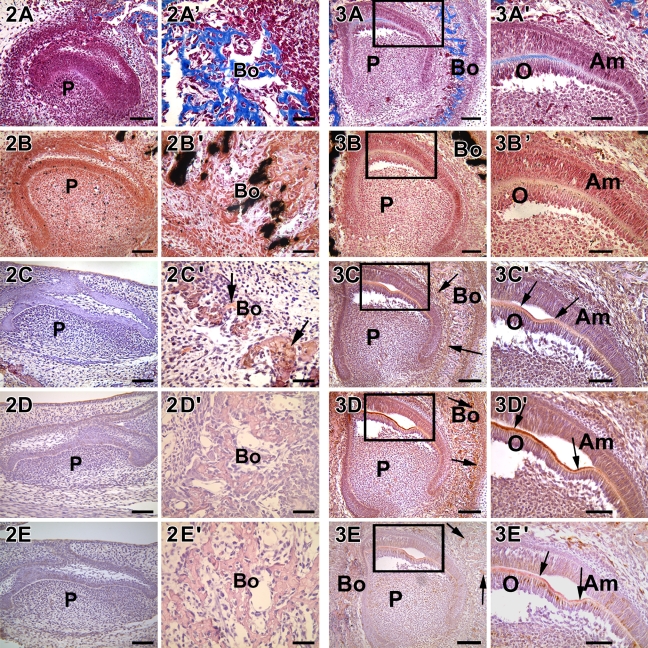
Figure 3.
Localization of collagen, mineral deposits, DMP1, DPP, and DSP in a postnatal 1-day-old mouse (P1). (A,A′) The incisor is at the late bell stage and trichrome Masson staining showed the deposition of the collagen matrix in the dentin and in the surrounding bone. (B,B′) Sparse deposits of calcified deposits were observed in dentin, whereas abundant calcified matrix was observed in the surrounding bone. (C,C′) DMP1, (D,D′) DPP, and (E,E′) DSP were localized to secretory odontoblasts near the cuspal region, which were actively synthesizing a mineralized matrix. Arrows indicate mineralized dentin and bone matrix. A′–E′ represent the boxed areas in A–E. Am, ameloblast; O, odontoblast; P, pulp. Bars: A–E = 20 μm; A′–E′ = 10 μm.

