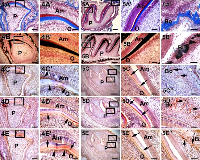
Figure 4.
Localization of collagen, mineral deposits, DMP1, DPP, and DSP in a postnatal 3-day-old mouse incisor. (A,A′ and B,B′) Progressive increase in collagen deposition and calcified matrix formation, respectively. DMP1 was localized at the mineralization front, which was between predentin and dentin, and in the odontoblasts (C,C′, arrow). (C) The surrounding alveolar bone also showed DMP1 localization. However, DMP1 was transiently expressed in the ameloblasts (C′, arrowhead). DPP and DSP were both localized at the mineralization front (D′,E′, arrow). DSP was highly expressed in the cytoplasm of the odontoblasts (E′, arrowhead). Both DPP and DSP were expressed in the ameloblasts. Bars: A–E = 20 μm; A′–E′ = 10 μm.

