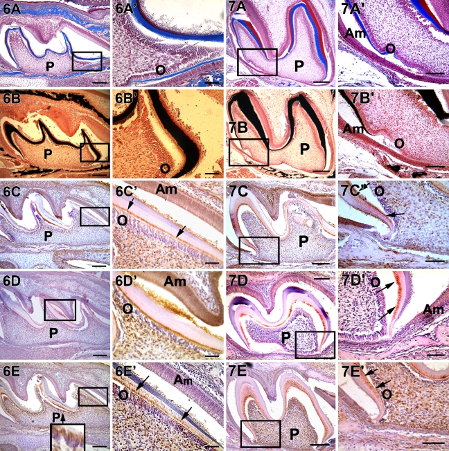
Figure 7.
Localization of collagen, mineral deposits, DMP1, DSP, and DPP in a postnatal 7 day mouse first molar. Deposition of collagen and mineralized matrix seen in A, B, A′, and B′. A′–E′ are higher magnification from the corresponding boxed portion from A–E. Arrows indicate localization of proteins at the mineralization front. Localization of DMP1, DPP, and DSP observed at the mineralization front and at the cervical loop region. DSP is localized in the bone at this stage of development. Bars: A–E = 20 μm; A′–E′ = 10 μm.

