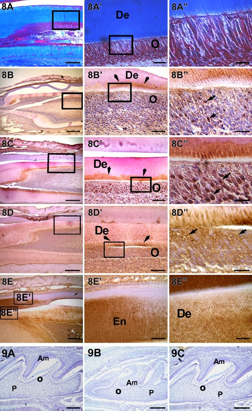
Figure 8.
Localization of collagen, DMP1, DSP, and DPP in a postnatal 20-day-old mouse incisor. Abundant collagenous matrix are localized in A,A′, and A″. DMP1 is localized at the mineralization front and also observed in the nucleus of differentiating odontoblasts (B,B′,B″). DPP is localized at the mineralization front and in the nucleus of the odontoblasts (C,C′,C″). DSP is observed in the dentinal tubules and around the plasma membrane of the odontoblasts (D,D′,D″). DSP is also localized in the early formed enamel matrix (E′) and in the dentin matrix (E″). Am, ameloblast; De, dentin; En, enamel; O, odontoblast; P, pulp. Bars: A–E = 20 μm; A′–E′ = 10 μm; A″–E″ = 2 μm.

