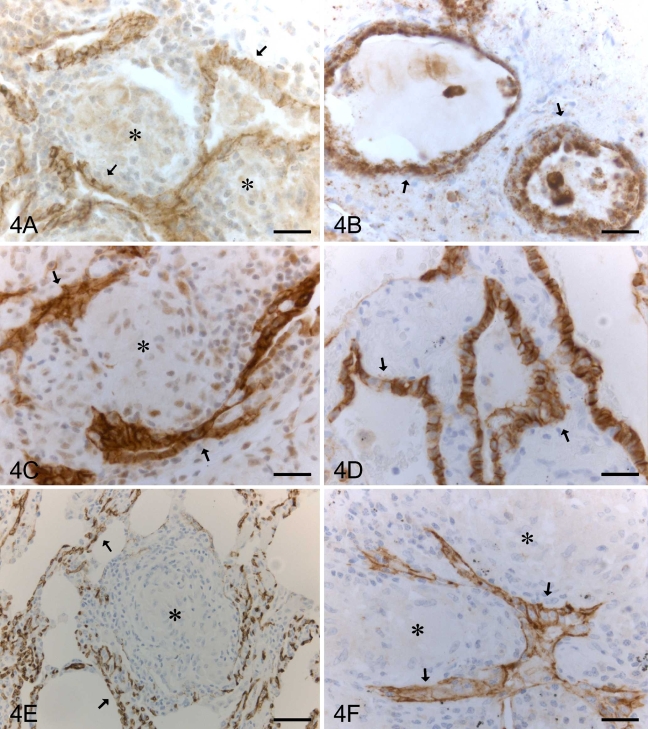
Figure 4.
IHC staining for claudin-1, -2, -3, -4, -5, and -7 in a patient with sarcoidosis. Metaplastic alveolar epithelium (arrows), which is often located beside granulomas (asterisks), is positive for claudin-1 (A), claudin-2 (B), claudin-3 (C), claudin-4 (D), and claudin-7 (F). (E) Endothelial cells of capillaries of alveolar walls are positive for claudin-5 (arrows) surrounding a granuloma (asterisk). Bar = 50 μm.

