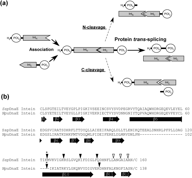
Figure 1. Protein trans-splicing and locations of the new split sites.
(a) Schematic representation of the protein trans-splicing process and two possible side reactions of N- and C-cleavage. Two fragments of the protein of interest (POI) can be ligated by protein trans-splicing reaction. (b) Sequence alignment of SspDnaE and NpuDnaE inteins. The locations of the experimentally tested split sites of SspDnaE and NpuDnaE inteins are indicated by inverse triangles on the top of the primary sequences. The asterisks above inverse triangles indicate the naturally occurring split site. Filled triangles indicate the split sites, where the split inteins retained protein trans-splicing activity. Open triangles indicate the split sites, where no protein trans-splicing activity could be detected. The location of the b-strands observed in the crystal structures of SspDnaE intein (PDB code 1ZDE) [35] and SspDnaB mini-intein (1MI8) [36] are indicated at the bottom of the sequences. The numbering for b-strands is adapted from SspDnaB mini-intein [36].

