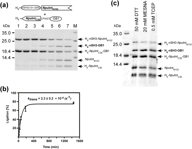
Figure 4. In vitro protein ligation of nSH3 and GB1 by the naturally occurring split NpuDnaE intein.
(a) Time course of the protein ligation of nSH3 and GB1 by naturally occurring split NpuDnaE intein in the presence of 50 mM DTT. Lane 1, 0 min after the mixing; lane 2, 3 min; lane 3, 10 min; lane 4, 30 min; lane 5, 1 hour; lane 6, 3 hours; lane 7, 22 hours. (b) Kinetic analysis of the protein ligation from the SDS-PAGE. (c) SDS-PAGE analysis of the ligation reaction after overnight incubation in the presence of different reducing agents.

