Figure 3.
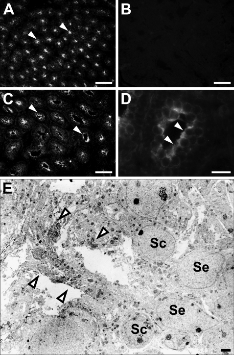
Immunohistochemistry showing the localization of radixin in the developing mouse testis. The frozen sections of 1-week (A,B) or 2-week (C–E) mouse testis were immunostained with rat monoclonal anti-radixin antibody (A,C–E) or normal rat IgG (B) and observed with fluorescence (A–D) or electron (E) microscope. (A) At 1 week, many seminiferous tubules display radixin immunoreactivity (arrowheads). (B) No immunoreactivity is present in any cells. (C) At 2 weeks, the immunoreactivity (arrowheads) is localized in the region facing the lumen of seminiferous tubules. (D) Higher magnification of the tubule in C. (E) Result of immunoelectron microscopy in the 2-week tubule. The immunoreactivity (arrowheads) is primarily localized in the apical cytoplasmic portions of Sertoli cells (Se). No immunoreactivity is present in spermatocytes (Sc). Bars: A–C = 50 μm; D = 25 μm; E = 1 μm.

