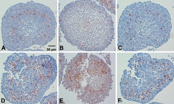
Figure 4.
IHC on paraffin sections of HT29 spheroids to active caspase-3 (A,D), active caspase-7 (B,E), or c-PARP (C,F) applied without treatment (A–C) or subjected to Foscan-PDT (D–F). Note the distribution of apoptotic cells at the periphery of necrotic area. Loss of cell cohesion was noticeable in spheroids subjected to Foscan-PDT (D–F). Harris' hematoxylin counterstain.

