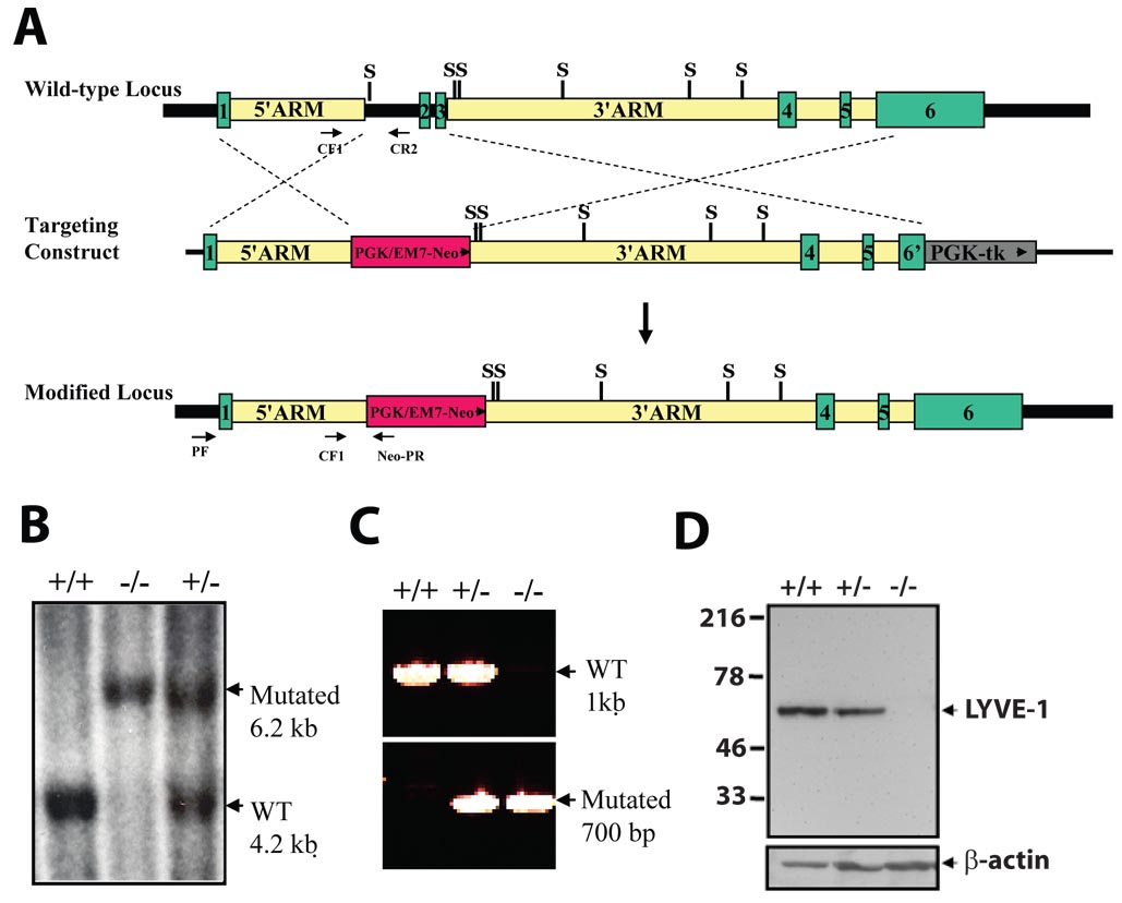
FIG. 1. Targeted inactivation of the mouse LYVE-1 gene.
(A) Schematic diagram of the LYVE-1 locus, the targeting construct and the mutant allele. Homologous recombination of the targeting vector introduces the neo gene downstream of the LYVE-1 promoter, deleting exons 2 and 3 of the LYVE-1 gene. Green boxes indicate the coding exons, whereas the dashed lines indicate the regions of identity between the LYVE-1 locus and the targeting vector. tk, selectable thymidine kinase gene; WT, wild-type. S, SspI. (B) Southern blot analysis of SspI-digested genomic DNA from targeted ES cells using an 856-bp 5’ external probe generated from PCR primers UF and UR. A 6.2-kb restriction fragment is diagnostic for the mutated allele, whereas a 4.2-kb fragment is generated from the wild-type allele. +/+, wild-type; +/−, heterozygous; −/−, homozygous mutant. (C) Genotyping by PCR analysis of genomic tail DNA. Primer pairs CF1/CR2 and CF1/neo-PR were used to detect the wild-type and mutant alleles, respectively. (D) Western blot analysis of proteins (20 µg) from lung tissue using LYVE-1 antibody detects a 60-kD band in wild-type and heterozygous samples, but not in homozygous mutant samples.

