Figure 3.
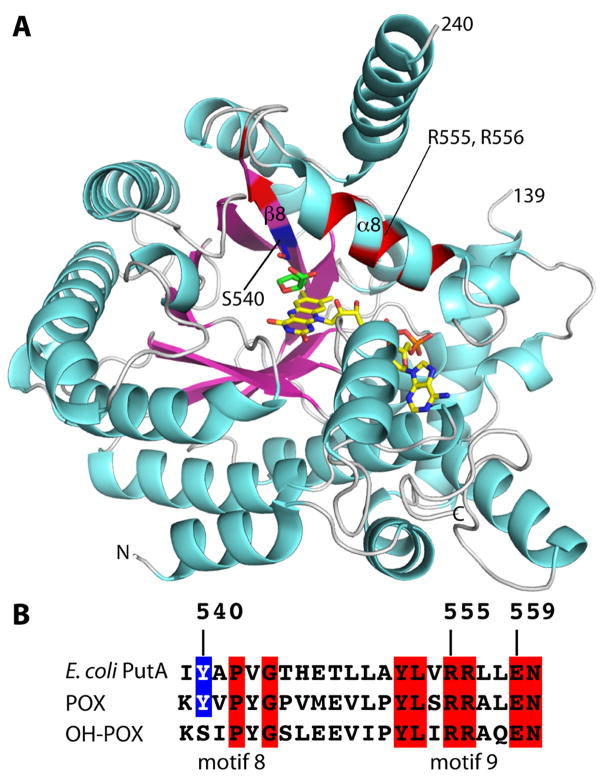
Overall structure of the PutA PRODH domain. (A) Ribbon drawing of PutA86-630 mutant Y540S complexed with THFA. FAD and THFA are drawn as sticks and colored yellow and green, respectively. The side chain of Ser540, which is located on β8, is shown in blue sticks. The locations of Arg555 and Arg556 of α8 are indicated. Residues highlighted in blue and red correspond to the sequence alignment shown in panel B. (B) A section of an amino acid sequence alignment of E. coli PutA, POX and OH-POX in the region of conserved motifs 8 and 9, which corresponds to secondary structural elements β8 and α8 as shown in panel A.

