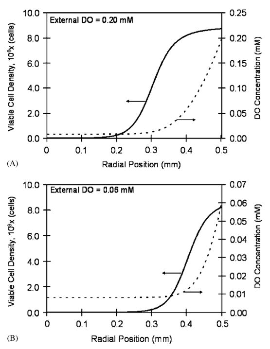
Fig. 1.

Steady-state viable cell density and DO profiles in a 1.0mm diameter bead at two external DO concentrations: (A) external DO equal to 0.20mM. At the periphery (radial position equal to 0.5mm), the cell density reaches 8.74 × 10⁸ cells/ml due to the high DO concentration (0.20mM). The cell density declines towards the center of the bead attaining its lowest value of 1.8 × 10⁵ cells/ml at the center where the DO concentration is 0.008mM. (B) External DO equal to 0.06mM. The maximum cell density at the periphery is 8.23 × 10⁸ cells/ml. At the center where the oxygen concentration is 0.008mM, the cell density declines to 5.0 × 10³ cells/ml.
