
Fig. 3.
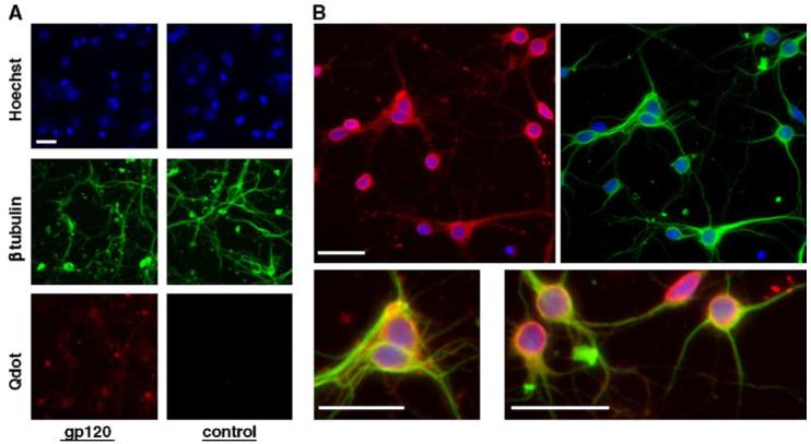
Binding of gp120 to primary cortical neurons. Pure cultures of rat cortical neurons (7 DIV) were incubated with either labeled gp120IIIB (400 pM) or with vehicle (in control cells), fixed and then stained with nanoprobes (Q-Dots). Neurons that bind gp120 appear in red. Hoechst 33342 (blue) was used for nuclear staining and β tubulin III immunostaining was used as a neuronal marker (green). Neurons were permeabilized (in order to perform the staining for the neuronal marker) after treatment with the nanoprobes. Therefore, only the gp120 bound to the plasma membrane was detected. Panel A shows both control and gp120-treated neurons, while in panel B enlarged images of gp120-treated cells are reported. The scale bar represents 20 μm.