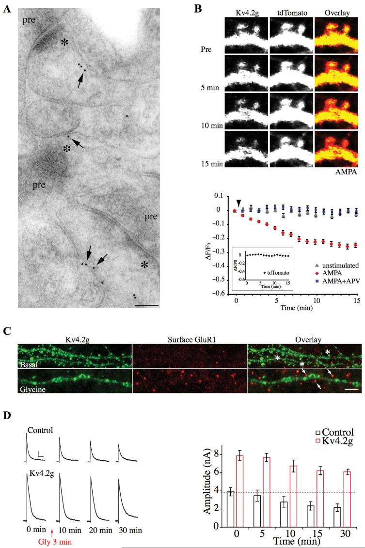
Fig. 1.
Synaptic Kv4.2 channels contribute to long-term potentiation. (A) Immunogold labeling for endogenous Kv4.2 showing its expression in CA1 spines. For electron micrographs, stars (*) indicate the postsynaptic density and “pre” indicates the presynaptic terminal. Scale bar: 100 nm. (B) Time-lapse images showing Kv4.2g fluorescent intensity decrease upon AMPA (50 μM) stimulation in spines of hippocampal neurons coexpressing Kv4.2g and tdTomato (top). Time course of averaged fractional fluorescent changes (ΔF/F0) of Kv4.2g in spines (bottom). AMPA stimulation resulted in a progressive decrease of Kv4.2g- specific fluorescent intensity in spines, with no significant change in tdTomato fluorescent intensity (inset). AMPA-mediated decreases in spine Kv4.2g fluorescence intensity were blocked by APV. (C) ChemLTP induced with brief glycine exposure, results in synaptic insertion of GluR1 but also Kv4.2g internalization. Stars = Kv4.2g-positive clusters; arrows = Kv4.2g-negative clusters. Scale bar: 8 μm. (D) Examples of the decrease in transient K+ currents during chemLTP. A-type K+ currents are decreased by Kv4.2 internalization during chemLTP. Scale bar: 1 nA, 100 ms. (Modified from Kim and others 2007.)

