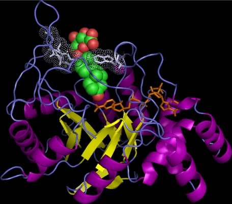
FIGURE 4.
The steroid binding site of AKR1C enzymes can accommodate conjugated steroids. A binding model of DhtG in AKR1C2 is shown. The details of the model building are given under “Experimental Procedures.” The (α/β)8-barrel structure of AKR1C2 is shown in ribbon representation with α-helices in purple and β-strands in yellow. NADP+, shown in stick representation in orange, binds in a highly conserved position. DhtG, shown in sphere representation with carbon in green and oxygen in red, binds in the steroid binding site that is formed by residues from several loops (in blue). The 3-keto group of the steroid is in close proximity with the nicotinamide ring of the cofactor for 3-ketosteroid reduction. The conjugate group of the steroid is accommodated in the wide opening at the entry point of the steroid binding pocket. The closest neighboring residues (in light blue) to the glucuronide group are shown in stick and dot representations. The figure was prepared using PyMOL.

