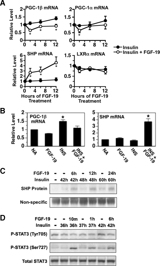
FIGURE 7.
Effect of FGF-19 on the expression of PGC-1β, PGC-1α, SHP, LXRα, and phosphorylated STAT3. A, primary hepatocyte cultures were prepared and incubated in Waymouth's medium containing insulin. At 36 h of incubation, FGF-19 (50 ng/ml) was added to the culture medium, and incubation was continued for 0, 2, 6, and 12 h. Cells were harvested, total RNA was isolated, and the abundance of the mRNAs encoding PGC-1β, PGC-1α, SHP, and LXRα was measured by quantitative real-time PCR. The level of mRNA in cells treated with insulin for 36 h and FGF-19 for 0 h was set at 1, and the other values were adjusted proportionately. Values are means ± four experiments. An asterisk indicates that the mean is significantly (p ≤ 0.05) different compared with that of cells treated with insulin for the same time period. B, primary hepatocyte cultures were prepared and incubated in Waymouth's medium. At 20 h of incubation, the medium was replaced with one of the same composition supplemented with or without FGF-19, insulin, or insulin plus FGF-19. After 24 h of treatment, total RNA was isolated, and the abundance of PGC-1β mRNA and SHP mRNA was measured by quantitative real-time PCR. The level of mRNA in cells incubated with no additions (NA) was set at 1, and the other values were adjusted proportionately. Values are means ± four experiments. An asterisk indicates that the mean is significantly (p ≤ 0.05) higher than that of any other treatment. Total cell extracts were prepared from hepatocytes treated as described in A. SHP protein (C) and phosphorylated STAT3 (Tyr705 and Ser727)(D) were measured by Western analysis. The data are representative of four independent experiments.

