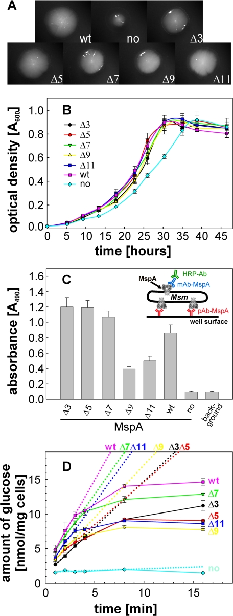
FIGURE 6.
Surface accessibility and in vivo function of MspA L6 deletion mutants. A, growth on agar plates and colony morphology. M. smegmatis ML16 complemented with MspA L6 deletion mutants were grown on HdB minimal medium supplemented with 1% glucose. After 5 days of incubation at 37 °C, pictures of representative colonies were taken at 12.5× magnification. B, growth in liquid medium. Triplicate cultures in beveled flasks were inoculated to an A600 of 0.02 in HdB liquid medium, 0.025% tyloxapol, and supplemented with 0.2% glucose. The optical density at 600 nm was recorded at the indicated time points during growth at 37 °C. C, surface accessibility of MspA mutants by whole cell capture ELISA. The experimental set-up is drawn as an insert. Wells of a Maxisorp 96-well microtiter plate were coated with an MspA antiserum to which ML16 cells expressing MspA were then “captured.” The anti-MspA mAb P2 was used for detection of surface-exposed MspA in combination with a secondary antibody conjugated with horseradish peroxidase. D, uptake of glucose. ML16 complemented with MspA mutants were grown to an A600 of ∼0.8 and incubated with a 20-μm mixture of nonlabeled and [14C]glucose. At the indicated time points, 1 ml of the cell suspensions were removed, filtered, and counted on a scintillation counter. The dotted lines represent the best fit for the first four time points (1-4 min). Uptake rates for wt, no MspA, Δ3, Δ5, Δ7, Δ9, and Δ11 were determined to be 2.5, 0.05, 1.3, 1.2, 2.0, 1.5, and 2.0 nmol/mg cells/min, respectively.

