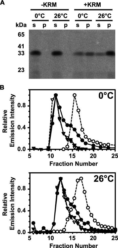
FIGURE 1.
RTA binds to ER microsomal membranes. 0.25 μm (A) 2.5 μm (B) RTA259-NBD were incubated in buffer H for 30 min on ice (0 °C) or at 26 °C with either microsomal membranes (+KRM; 20-40 eq) or an equal amount of buffer without microsomes (-KRM). Free RTA259-NBD was then separated from KRM-bound RTA259-NBD either by sedimentation (A) or by gel filtration chromatography (B). A, following sedimentation, the supernatant (s) and the microsomal pellet (p) were analyzed by SDS-PAGE. NBD-labeled proteins were visualized using a fluorescence imager. B, following mixing, free and KRM-bound RTA259-NBD were separated by gel filtration chromatography in buffer H at 4 °C using a Sepharose CL-2B column (8 × 0.5-cm inner diameter). Each fraction was scanned for the presence of RTA259-NBD (•; λex = 468 nm; λem = 530 nm) and KRMs (▾; λex = 405 nm; λem = 420 nm). As controls, only RTA259-NBD (○) or only KRMs (▿) were run and analyzed separately.

