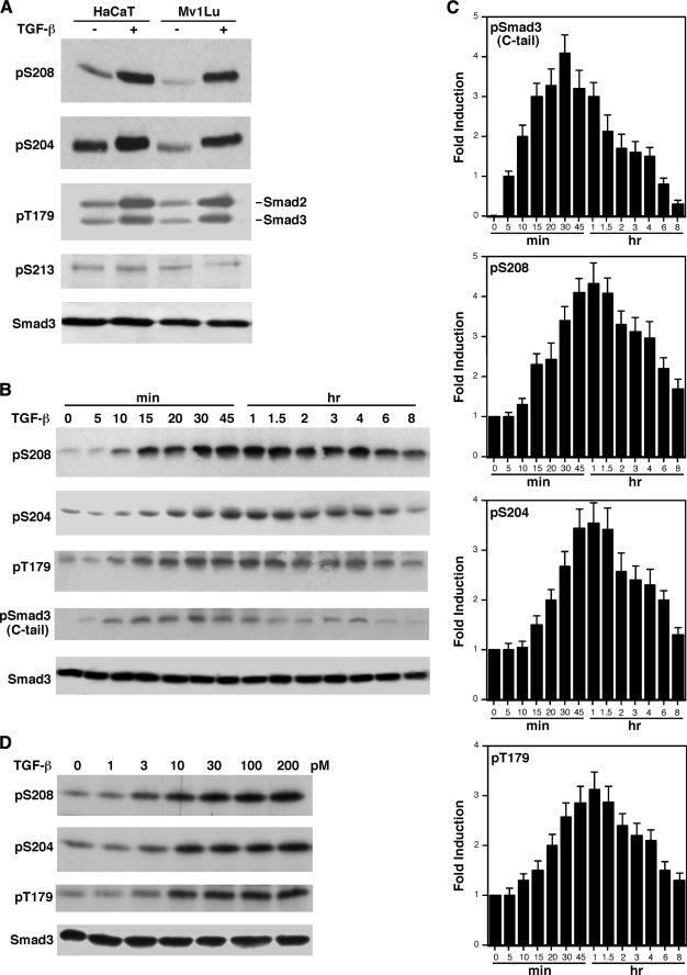
FIGURE 1.
TGF-β induces rapid phosphorylation of Smad3 linker sites. A, phosphorylation of Ser208, Ser204, and Thr179 in the Smad3 linker region is significantly increased by TGF-β. Mv1Lu and HaCaT cells were treated with 300 pm TGF-β for 1 h. Phosphorylation of Ser208, Ser204, Thr179, and Ser213 sites were detected by immunoblots using specific phosphopeptide antibodies. B, time course of TGF-β-inducible phosphorylation of Smad3. Mv1Lu cells were treated with 300 pm TGF-β for the indicated periods of time. Phosphorylation of Ser208, Ser204, and Thr179 as well as the C-tail were analyzed by specific phosphopeptide antibodies. Smad3 protein levels were also examined by immunoblot as a control. C, the average of four independent time course experiments from B. The error bars indicate the standard deviation. D, TGF-β dose curve for Ser208, Ser204, and Thr179 phosphorylation. Mv1Lu cells were treated for 1 h with the indicated concentrations of TGF-β. Phosphorylation of Ser208, Ser204, and Thr179 and Smad3 expression levels were analyzed by immunoblots.

